")

Infection Immunology

News
- 05.03.2026. A study conducted with the University of Freiburg shows for the first time that resident Kupffer cells – and not immigrated monocytes – functionally reprogram themselves in the granuloma core and control the anti-mycobacterial response.
- 20.11.2025. The complement factors C1q and MBL recognise tuberculosis bacteria and activate the complement system, with C1q playing the main role, without significantly impairing the viability of the bacteria.
- 02.06.2025. A mouse-to-human translational model accurately predicted the efficacy of BTZ-043 in humans.
- 18.02.2025. Novel antibiotic BTZ-043 also reaches tuberculosis bacteria hiding in dead lung tissue -> Press release
- 30.01.2025. Molecular Bacterial Load Assay (MBLA): New method can quickly and accurately predict the effectiveness of tuberculosis treatment -> Press release
- 01.10.2024. A new BDM-based tuberculosis treatment is more effective than the standard treatment and could significantly shorten the duration of therapy.
Tuberculosis (TB) is a chronic infectious disease of the lungs caused by Mycobacterium tuberculosis. After transmission via aerosols, the pathogen enters the alveoli, where it is taken up by immune cells and enclosed in so-called granulomas. In most cases, the immune system is able to control the infection, resulting in latent TB. In approximately 5–10% of infected individuals, this immune barrier fails, exacerbated by factors such as malnutrition, diabetes or HIV. This leads to progressive lung damage and active TB with symptoms such as coughing, weight loss and night sweats.
Despite antibiotics, TB treatment remains challenging. Drug-sensitive TB is treated for six months with a combination of rifampicin, isoniazid, pyrazinamide and ethambutol. The treatment of drug-resistant TB (MDR/XDR-TB), for which standard drugs are ineffective, is more difficult. In this case, lengthy combinations of new active substances such as bedaquiline, pretomanid and linezolid are used, but these are often toxic and do not always lead to a cure. Even after successful antimicrobial therapy, long-term lung damage may remain. This so-called post-tuberculous lung disease (PTLD) is characterised by irreversible structural changes, chronic airway obstruction and impaired lung function, which can have a lasting impact on quality of life and mortality.
The granuloma is the central immunological structure of TB and the site where it is determined whether an infection is controlled or progresses to disease. In a protective granuloma, the immune response is functionally organised so that long-lived immune cells effectively contain the bacterium while limiting tissue damage. In severe cases, however, damaging granulomas develop, characterised by necrosis, fibrosis and an immunosuppressive environment, which promote the survival of the pathogen. At the same time, the granuloma is the decisive site of action for antibiotics, whose effectiveness depends largely on the structure, immune composition and accessibility of these lesions.
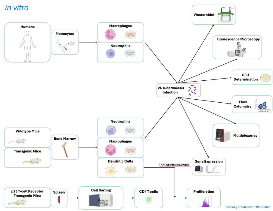
The Infection Immunology research group therefore aims to elucidate the immunological protective and damaging mechanisms of TB in granulomatous lung lesions in order to identify new therapeutic targets against active, resistant and persistent infections. Using a translational approach, the group combines preclinical drug development and immunomodulation in experimental mouse models to enable more effective therapies and prevent long-term sequelae such as PTLD.
Protective granulomas
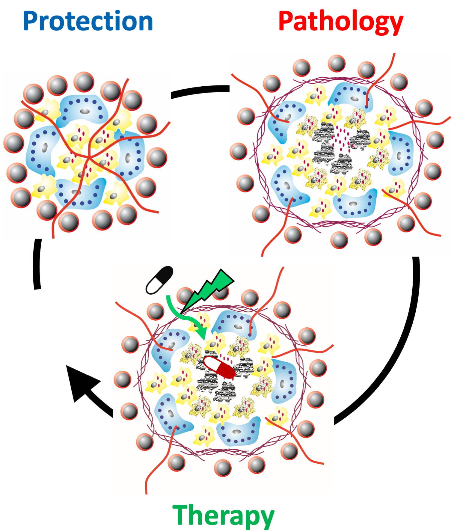
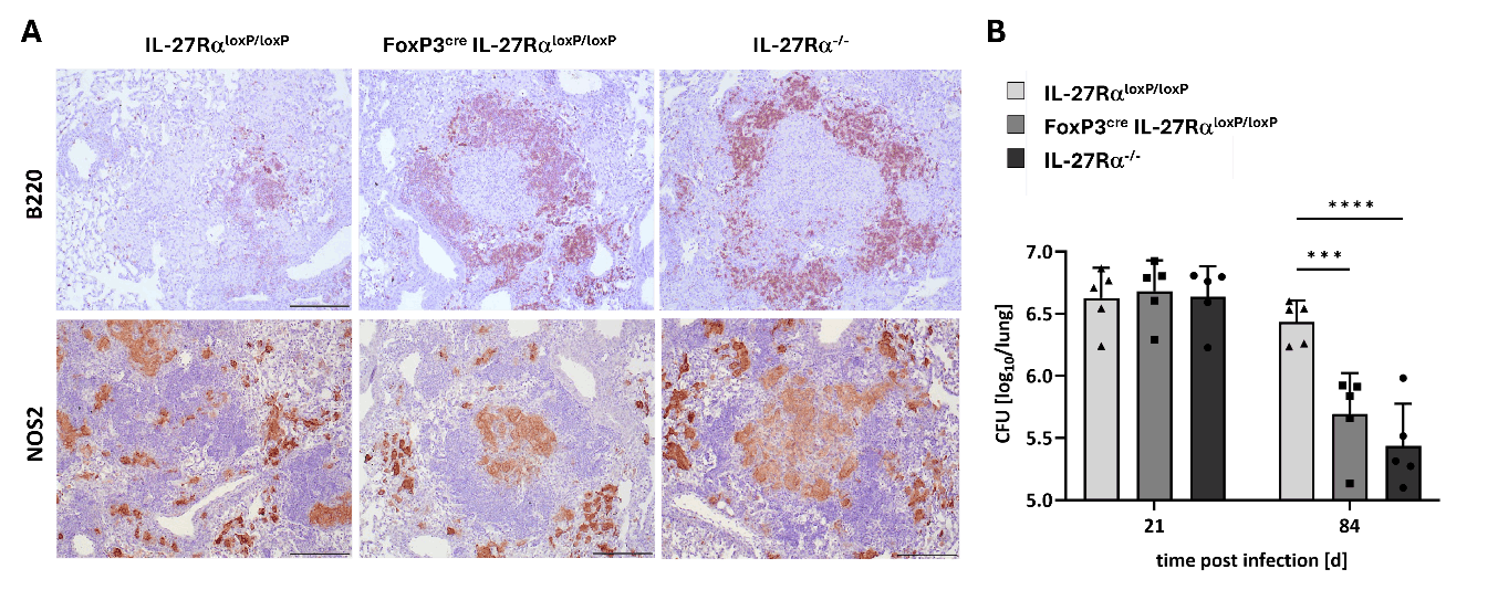
Protective granulomas can suppress an infection with Mycobacterium tuberculosis over the long term. They are functionally organised and contain long-lived immune cells that enable bacterial control. This form of protective immune organisation is primarily supported by pro-inflammatory T helper cells (Th) of the Th1 and Th17 types, which provide specific signals to activate infected macrophages and enhance antibacterial mechanisms in the lungs. At the same time, their activity is regulated by inhibitory cytokines to limit tissue damage. In particular, the pro-inflammatory cytokine interleukin (IL)-17, which is mainly produced by Th17 cells and gamma-delta T cells, plays an important role during granuloma development. In order to gain a more detailed understanding of the cytokine-mediated control mechanisms for establishing and maintaining protective granulomas, the Infection Immunology research group is interested in the differential roles of cytokines and cytokine receptors associated with the ubiquitous receptor subunit gp130. In this context, we were able to show that the gp130-dependent cytokine IL-6 plays a minor role in the development of IL-17A-expressing Th17 cells during experimental tuberculosis (TB), in contrast to its key role in the induction of Th17 immune responses in other inflammatory diseases. Accordingly, the absence of IL-6 or gp130 on T cells has little effect on the containment of the infection. In contrast, a global deficiency of the cytokine receptor subunit IL-27Rα in M. tuberculosis-infected mice leads to reduced bacterial load, but also to increased immunopathology. IL-27Rα together with gp130 forms the receptor of the heterodimeric cytokine IL-27. The enhanced containment of mycobacteria in IL-27Rα-deficient mice is accompanied by the formation of highly structured protective granulomas, which consist of a core of infected macrophages surrounded by a rim of protective T and B cells. By analysing the course of M. tuberculosis infection in IL-27Rα x IL-17A double-deficient mice, we were able to further demonstrate that the protective effect of IL-27Rα deficiency is dependent on the correlating increased expression of the cytokine IL-17A. IL-27 thus appears to limit the development of a protective immune response against M. tuberculosis, partly through the inhibition of IL-17A. At the same time, however, the suppression of IL-17A expression by IL-27 also leads to a reduction in immunopathological consequences. Most recently, we were able to demonstrate in mice with a cell type-specific deficiency of IL-27Rα-mediated signalling in regulatory T cells that the inhibitory effect of IL-27 on the development of protective granulomas is partly due to direct IL-27-mediated induction of these immunosuppressive cells (Fig. 1). Overall, this work provides crucial foundations for the further development of adjuvant therapeutic strategies with the aim of optimising the architecture of granulomatous lung lesions through targeted immunomodulation.
Literature: Ritter & Behrends et al. 2022 Cells.; Ritter et al. 2022 Front Immunol.; Ritter et al. 2021 J Mol Med (Berl); Ritter & Sodenkamp et al. 2020 Cells.; Ritter et al. 2020 Cells.; Erdmann & Behrends et al. 2018 Mucosal Immunol.; Sodenkamp et al. 2012 Immunobiology.; Sodenkamp et al. 2011 Eur J Cell Biol.; Hölscher et al. 2005 J Immunol.
The complement system acts as a first line of defence against pathogens and is a central component of the innate immune response. However, the relevance of the complement system in TB has hardly been investigated so far, and it is not known whether the protein cascade contributes to increased protection or damage. Regarding the interaction of complement proteins with mycobacteria, we have recently shown that different clinical isolates of the M. tuberculosis complex bind complement components and activate the cascade. In addition, our initial in vivo studies suggest that the anaphylatoxin-mediated inflammatory response influences the formation of TB granulomas. Future studies will aim to characterize the complement-mediated immune response and pathophysiology in more detail with the aim of validating complement modulation as a potential adjunct therapy for TB.
Literature: Duque et al. 2025 J Immunol; Krusch 2018 Promotion.
Damaging granulomas
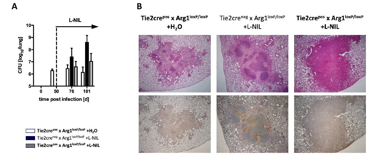
Damaging granulomas are caused by a misdirected immune response in which protective mechanisms for controlling pathogens are converted into tissue-destructive processes. The central histopathological feature of human TB, central granuloma necrosis following infection with M. tuberculosis, does not typically occur in standard laboratory mice. Based on our investigations in TB patients, the Infection Immunology research group described a functional SNP in the IL4RA gene that is associated with enhanced signal transduction and more severe pulmonary pathology. This led us to the hypothesise that a Th2-dominated immune response mediated by IL-4Rα contributes significantly to TB pathology. Since conventional mice do not develop a corresponding Th2 immune response after infection with M. tuberculosis, we infected IL-13-overexpressing mice with M. tuberculosis. In these animals, the IL-13-driven Th2 immune response via the IL-4Rα signalling pathway led to the formation of central necrotising granulomas, which are the essential histopathological feature of human post-primary TB. This provides the Infection Immunology research group with an experimental model that enables the targeted analysis of downstream immunological and metabolic mechanisms of TB-associated tissue pathology. In this context, there is pronounced alternative activation of macrophages with strong arginase-1 expression, lipid accumulation and formation of a hypoxic necrotic granuloma core surrounded by a collagen capsule. These arginase-1-positive foam macrophages promote both pathological tissue remodelling and bacterial persistence within the granuloma. In addition, we were able to show that damaging granuloma necrosis can also be induced independently of IL-13 when the NOS2-dependent antimicrobial metabolic pathway is pharmacologically blocked by the inhibitor L-NIL. In standard C57BL/6 mice, this targeted inhibition leads to a shift in L-arginine metabolism towards arginase-1 activity and the formation of centrally necrotic granulomas with fibrotic collagen rings. Crucially, this pathology is completely eliminated in mice with macrophage-specific knockout of arginase-1, even though NOS2 inhibition remains intact (Fig. 2). The work of the Infection Immunology research group thus demonstrates that arginase-1-dependent mechanisms are a central, common endpoint of damaging granuloma formation and contribute significantly to the pathology, resistance to therapy and progression of TB.
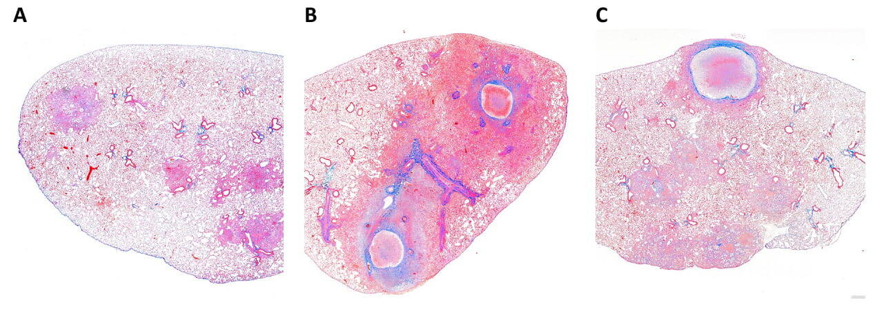
Unlike standard inbred strains such as C57BL/6, both IL-13tg mice and C3HeJ/FeB mice develop the granulomatous pathology typical of human TB, with central necrosis, fibrosis and hypoxic microenvironments (Fig. 3). Both models thus offer the opportunity to identify common pathogenetic features of these pronounced lesions and, on this basis, to uncover previously unknown downstream mechanisms of TB pathology and to evaluate the efficacy of antibiotics under pathophysiologically relevant conditions in a preclinical setting.
Literature: Walter et al. 2022 Antimicrob Agents Chemother; Brandenburg et al. 2021 J Clin Invest; Lösslein et al. 2021 Nat Commun; Schmok et al. 2017 Front Immunol; Herrtwich et al. 2016 Cell; Hölscher et al. 2016 Mediators Inflamm; Heitmann et al. 2014 J Pathol; Schreiber et al. 2009 J Immunol.
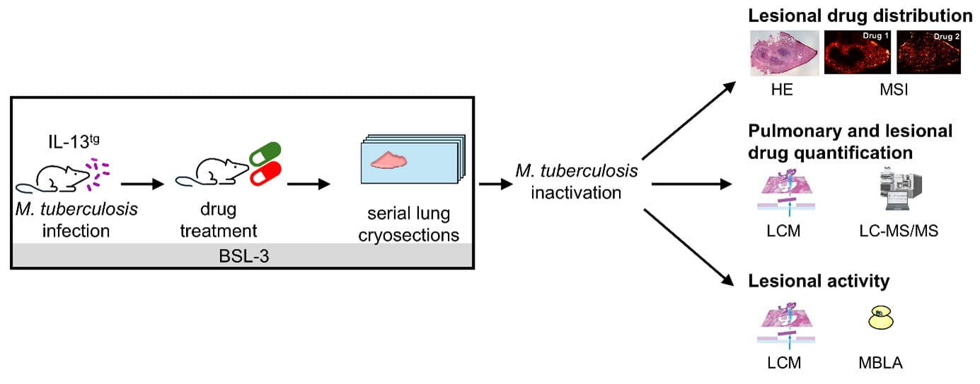
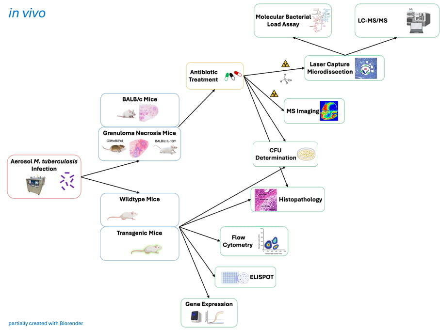
Evaluation of antibiotics in preclinical mouse models
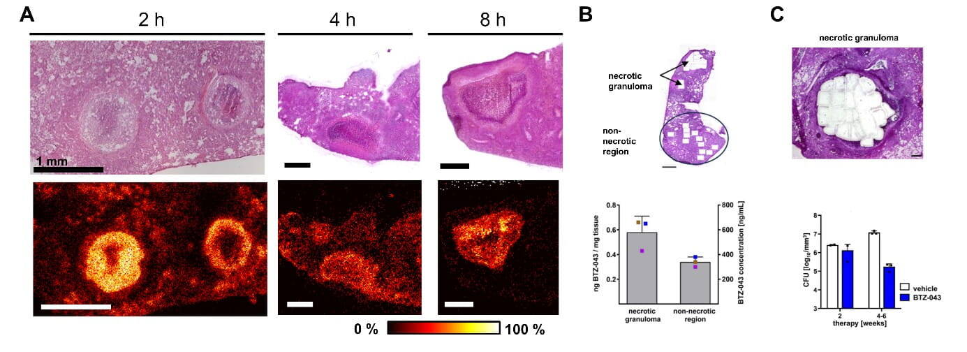
Antibiotic-resistant strains of M. tuberculosis are a major threat to global control of TB, which is why new anti-TB drugs are urgently needed. Drug development against multidrug-resistant TB (MDR-TB) requires preclinical validation in advanced animal models that reflect the pathophysiological aspects of human TB, since the complex structure of centrally necrotizing granulomas favours mycobacterial survival but hampers the penetration of many antibiotics. Therefore, the Infection Immunology research group, in collaboration with other scientists within the German Centre for Infection Research (DZIF), has established a powerful preclinical infrastructure for the improved evaluation of novel anti-mycobacterial compounds, which is also integrated into international networks (ERA4TB and UNITE4TB). A fundamental pillar of this preclinical platform are advanced mouse models (IL-13tg, C3HeB/FeJ), that develop a human-like granuloma pathology with a mycobacteria harbouring necrotic core and fibrotic encapsulation. For a comprehensive in vivo validation of novel drug candidates, we combine these advanced mouse models with state-of-the-art methods for drug detection, such as mass spectrometry imaging (MSI, collaboration with Andreas Römpp, University of Bayreuth) or laser capture microdissection (LCM) coupled with LC-MS/MS (collaboration with Dominik Schwudke, Research Centre Borstel). By combining LCM with molecular methods for the detection of M. tuberculosis (molecular bacterial load Assay, MBLA) we determine the pathogen burden within necrotic granulomas (Fig. 4). This innovative approach was used for the comprehensive validation of the novel antibiotic BTZ-043 revealing that BTZ-043 fully penetrates centrally necrotising granulomas, at concentrations manifold above its in vitro minimum inhibitory concentration (1 ng/ml) and that BTZ-043 exerts its antibacterial activity within necrotic lesions(Fig. 5). Based on these properties, BTZ-043 has the potential to complement existing TB antibiotics and may contribute to future regimens to shorten treatment duration, prevent reactivation and avoid the development of drug resistance. The efficacy of BTZ-043-based combination therapies are currently being evaluated in clinical trials (UNITE4TB and PanACEA).
In addition, we use the BALB/c standard mouse model in our preclinical infrastructure to analyse the anti-mycobacterial activity of novel drug candidates during acute and chronic M. tuberculosis infection. We also investigate TB reactivation in the BALB/c relapse model and validate the sterilising activity of novel antibiotic combinations.
In summary, our preclinical infrastructure supports the development of better treatment options for TB patients and contributes to the prevention of antimicrobial resistance.
Literature: Römpp et al. 2025 Nat Commun; Ngara et al. 2025 J Infect Dis; Walter & te Brake et al. 2024 J Antimicrob Chemother; Joch et al 2024 Eur J Med Chem; Walter et al. 2022 Antimicrob Agents Chemother; Kokesch-Himmelreich et. al 2022 Anal Chem; Treu et al. 2020 J Am Soc Mass Spectrom.
Immunomodulation in granuloma as a therapeutic strategy
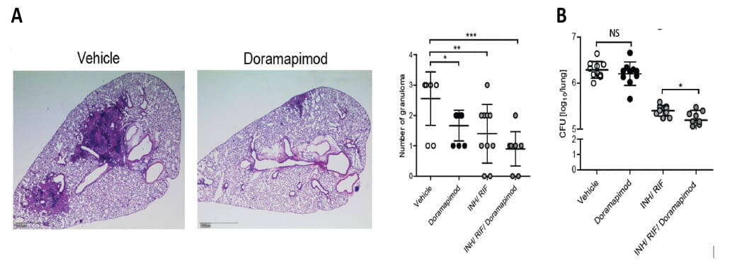
In a comprehensive research project, the Infection Immunology research group is investigating how the formation of necrotic granuloma lesions can be therapeutically influenced by targeted immunomodulation in IL-13tg and C3HeB/FeJ mice. By inhibiting key immunometabolic and inflammatory signalling pathways – including Arg-1, p38MAPK, NLRP3, ACC2, IL-27Rα and anaphylatoxin receptors – the inflammatory and metabolic environment within the granulomas is specifically altered, thereby attenuating necrotic processes and stabilising tissue architecture. This modulation not only leads to improved local immune balance but also facilitates the penetration of antibiotics into pathologically altered lesions, thereby increasing their effectiveness under disease-relevant conditions. While inhibition of these targets in C57BL/6 mice already leads to a significant reduction in pulmonary inflammation (Fig. 6), our IL-13tg and C3HeB/FeJ models will enable the investigation of these approaches in complex, human-like granulomas. The project thus combines the mechanistic elucidation of damaging immune responses with the development of adjuvant, immunomodulatory therapeutic strategies to improve existing antibiotic regimens.
Literature: Theobald et al. 2021 Cell Death Discov; Brandenburg et al. 2021 J Clin Invest; Hölscher et al. 2020 Sci Rep; Gräb et al. 2019 Nat Commun
Post-tuberculosis lung disease
Approximately one half of successfully treated TB patients continue to suffer from persistent lung impairment after the end of treatment. The term "post-tuberculous lung disease" (PTLD) covers a broad spectrum of abnormalities that can affect the airways, the lung parenchyma, the pulmonary vascular system and the pleura, including bronchiectasis, cavitations and pulmonary fibrotic changes. At the immunological level, the development of PTLD is typically accompanied by a shift towards a Th2-dominated immune response, accompanied by the alternative activation of macrophages, which contribute to the progression of the disease. Increased expression of Th2 cytokines also promotes the formation of collagen deposits and the proliferation of fibroblasts, leading to pulmonary fibrosis and scarring. Since the IL-13tg mouse model is not only characterised by the formation of human-like central necrotising granulomas after infection with M. tuberculosis but also induces key features of PTLD through its IL-13 overproduction, these animals represent an excellent tool for studying this disease. The planned project will focus both on the detailed analysis of the immunopathological factors associated with the various forms of the disease and on functional analyses of the lungs, with the ultimate aim of developing approaches to prevent post-infectious lung damage through the targeted modulation of identified target structures.
The development of a vaccine against rickettsioses
Rickettsioses are emerging febrile, potentially fatal infectious diseases caused by small intracellular bacteria (rickettsiae). Rickettsial infections are highly prevalent in developing countries and a serious global health threat. Within the past decades, rickettsial infections have been occurring around the world with increasing frequency and geographic distribution. Since the bacteria only respond to very few antibiotics, treatment with wrong antibiotics or delayed treatment due to a lack of diagnosis or misdiagnosis often leads to severe disease courses. It is problematic that there is evidence of the development of antibiotic resistance. In addition, some rickettsiae species can persist and cause recurrent diseases regardless of antibiotic treatment. Finally, certain rickettsial species are classified as potential biological weapons. For these reasons, prophylactic vaccines against the infection with these bacteria are urgently needed. The aim of this project is to gain deeper insight into protective immune mechanisms and the identification of immunogenic determinants of rickettsial pathogens that can serve as a vaccine. Rickettsia typhi, the causative agent of endemic typhus, is used as a model organism for these studies that are funded by the German Research Foundation (DFG, No. OS583) and conducted by Dr. Anke Osterloh.
Awarded with the Memento Award for Neglected Diseases 2020
Literature: Osterloh. 2022 Vaccines (Basel); Osterloh. 2021 Vaccines (Basel); Rauch et al. 2021 PLoS One; Osterloh. 2020 PLoS Negl Trop Dis; Kopf et al. 2018 Antimicrob Agents Chemother
- BMFTR; DZIF TTU-TB 02.717 Infrastructure „Myco Drug and Trials“ – 2030
- BMFTR; DZIF TTU-TB 02.823 Project „Innovative Strategies for Tuberculosis Treatment: Discovery, Development, and Preclinical Evaluation of Novel Antimicro-bials and Host-Directed Therapies” – 2028
- BMFTR; Application of artificial intelligence (AI) in drug discovery "AI-driven fragment-based design of mycobacterial thioredoxin reductase inhibitors as new anti-tuberculosis drugs (KI-FIRE-TB)“ – 2028
- EU Horizon 2020; IMI “ERA4TB European Regimen Accelerator for Tuberculosis” – 2026
- DFG; OS 583/1-3 “Identification of immunodominant CD4+ T cell antigens as vaccine candidates for protection against infection with Rickettsia typhi and characterisation of the Rickettsia typhi-specific CD8+ T cell response“ – 2027
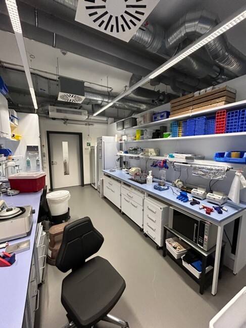
A new start after 25 years
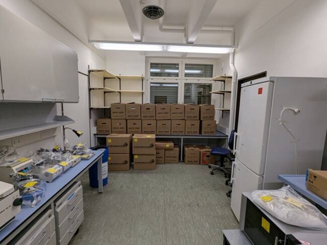
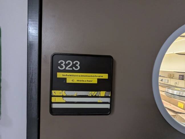
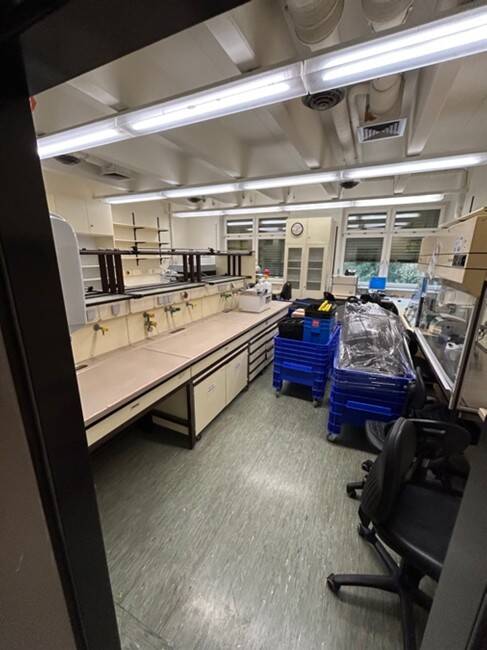
After a quarter of a century, it's time for our research group to pack boxes and discover something new. Our lab has moved to a new building – a step that was not without a touch of sadness. After all, over many years, not only experiments but also ideas, discussions and many a success story have been created in this place. This makes the joy of the new beginning all the greater: in a modern, bright laboratory environment, new opportunities for research, exchange and innovation are opening up for us. The fresh space and contemporary equipment are already creating a palpable sense of optimism. We hope that the BSL3 lab and the animal facility will soon follow and also be able to move into the new building. Until then, we are looking forward to everything that lies ahead – and to writing the next chapters of our research in a new location.


Congratulations, Alejandro, on successfully defending your doctoral thesis!
As the only one working with human cells in an environment full of mouse research, you have demonstrated perseverance and humour – sometimes surely feeling like you were in a zoo. Scientifically, you have brought new attention to the complement system as a classic player in immunology in the context of tuberculosis. A strong thesis with a clear message.
All the best for your future endeavours!
¡Enhorabuena, Alejandro, por la exitosa defensa de tu tesis doctoral!
Como el único que trabajó con células humanas en un entorno dominado por la investigación en ratones, demostraste perseverancia y sentido del humor — en algún momento seguro que se sintió un poco como estar en un zoológico. Desde el punto de vista científico, has conseguido devolver al sistema del complemento, un actor clásico de la inmunología, un lugar destacado en la investigación sobre la tuberculosis. Un trabajo excelente con un mensaje claro.
¡Mucho éxito en lo que venga ahora!

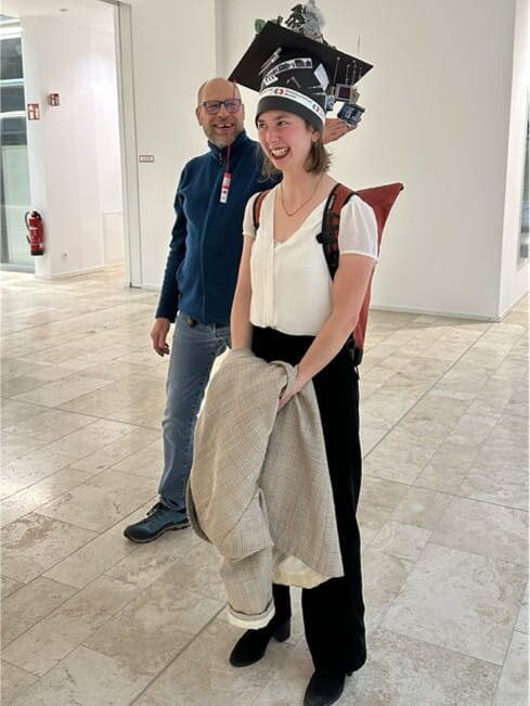
Congratulations, Sofia! 🎉
We would like to congratulate Dr Sofia Scheele on the successful defence of her doctoral thesis. With great dedication, she has established laser capture microdissection (LCM) at our institute and thus set a real milestone for the research group. Dear Sofia, we are proud of you – and you will always remain part of our family.
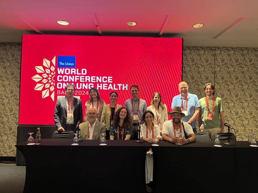
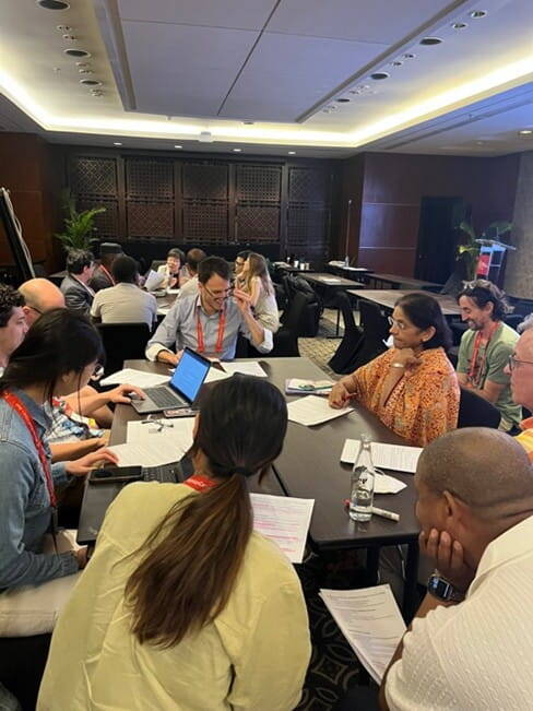
International ERA4TB workshop at the Union World Conference in Bali
As part of the Union World Conference in Bali, Christoph Hölscher and his colleagues from the ERA4TB consortium organised the workshop "TB drug regimen development school: advanced preclinical and translational tools with the public-private ERA4TB consortium hub". The workshop brought together participants from all over the world and provided a lively platform for exchange on modern preclinical and translational approaches in TB therapy development. The great international response and the lively discussions made the event a complete success.
Congratulations to Dr. Anna Baumann on her successful thesis defence!
Her dissertation provides important new insights into the role of arginase-1 as a susceptibility factor for Tuberculosis, above all, because "there was a worm in it" (one aspect of the work was M. tuberculosis – helminth co-infection). We warmly congratulate her on this success.

2026
Neuber, J, Lohrmann, F, Wald, S, Göçer, M, Lösslein, AK, Obwegs, D, Rogg, M, Gres, V, Kolter, J, Goldmann, T, Walter, K, Hölscher, C, Schell, C, Preissl, S, Sagar & Henneke, P 2026, 'Kupffer cell plasticity regulates hepatic immunity in mycobacterial infection: Kupffer cell plasticity', Journal of hepatology. https://doi.org/10.1016/j.jhep.2026.02.024
2025
Duque-Villegas, MA, Götz, MP, Rousseau, E, Homolka, S, Niemann, S, Garred, P, Hölscher, C, Walter, K* & Rosbjerg, A* 2025, 'C1q and mannose-binding lectin binding and complement activation across genetically diverse Mycobacterium tuberculosis complex strains', JOURNAL OF IMMUNOLOGY. https://doi.org/10.1093/jimmun/vkaf294 *authors contributed equally
Ngara, B, Flori, L, van Wijk, RC, Ernest, JP, Tyagi, S, Soni, H, Hölscher, C, Walter, K, Dreisbach, J, Hoelscher, M, Nuermberger, EL & Savic, R 2025, 'Translational modeling of BTZ-043 in predicting phase IIA efficacy and evaluating drug-drug interactions with BPaL in murine models', JOURNAL OF INFECTIOUS DISEASES. https://doi.org/10.1093/infdis/jiaf088
Römpp, A, Treu, A, Kokesch-Himmelreich, J, Marwitz, F, Dreisbach, J, Aboutara, N, Hillemann, D, Garrelts, M, Converse, PJ, Tyagi, S, Gerbach, S, Gyr, L, Lemm, A-K, Volz, J, Hölscher, A, Gröschel, L, Stemp, E-M, Heinrich, N, Kloss, F, Nuermberger, EL, Schwudke, D, Hoelscher, M, Hölscher, C & Walter, K 2025, 'The clinical-stage drug BTZ-043 accumulates in murine tuberculosis lesions and efficiently acts against Mycobacterium tuberculosis', Nature communications, Jg. 16, Nr. 1, S. 826. https://doi.org/10.1038/s41467-025-56146-9
2024
Götz, MP, Duque Villegas, MA, Fageräng, B, Kerfin, A, Skjoedt, M-O, Garred, P & Rosbjerg, A 2024, 'Transient Binding Dynamics of Complement System Pattern Recognition Molecules on Pathogens', JOURNAL OF IMMUNOLOGY, Jg. 212, Nr. 9, S. 1493-1503. https://doi.org/10.4049/jimmunol.2300768
Neumann, M, Reimann, M, Chesov, D, Popa, C, Dragomir, A, Popescu, O, Munteanu, R, Hölscher, A, Honeyborne, I, Heyckendorf, J, Lange, C, Hölscher, C* & Kalsdorf, B* 2024, 'The Molecular Bacterial Load Assay predicts treatment responses in patients with pre-XDR/XDR-tuberculosis more accurately than GeneXpert Ultra MTB/Rif', Journal of infection, S. 106399. https://doi.org/10.1016/j.jinf.2024.106399 *authors contributed equally
Walter, K, Te Brake, LHM, Lemm, A-K, Hoelscher, M, Svensson, EM, Hölscher, C* & Heinrich, N* 2024, 'Investigating the treatment shortening potential of a combination of bedaquiline, delamanid and moxifloxacin with and without sutezolid, in a murine tuberculosis model with confirmed drug exposures', JOURNAL OF ANTIMICROBIAL CHEMOTHERAPY. https://doi.org/10.1093/jac/dkae266 *authors contributed equally
2023
Joch, M, Wojtas, KP, Torres-Gómez, H, Li, Y, Meyer, F, Straßburger, M, Kerndl, V, Dahse, H-M, Hertweck, C, Hoffmann, H, Görls, H, Walter, K, Hölscher, C & Kloss, F 2024, 'Whole cell hydride Meisenheimer complex biotransformation guided optimization of antimycobacterial benzothiazinones', EUROPEAN JOURNAL OF MEDICINAL CHEMISTRY , Jg. 264, S. 116023. https://doi.org/10.1016/j.ejmech.2023.116023
2022
Brandenburg, J, Heyckendorf, J, Marwitz, F, Zehethofer, N, Linnemann, L, Gisch, N, Karaköse, H, Reimann, M, Kranzer, K, Kalsdorf, B, Sanchez-Carballo, P, Weinkauf, M, Scholz, V, Malm, S, Homolka, S, Gaede, KI, Herzmann, C, Schaible, UE, Hölscher, C, Reiling, N & Schwudke, D 2022, 'Tuberculostearic Acid-Containing Phosphatidylinositols as Markers of Bacterial Burden in Tuberculosis', ACS infectious diseases, Jg. 8, Nr. 7, S. 1303-1315. https://doi.org/10.1021/acsinfecdis.2c00075
Kokesch-Himmelreich, J, Treu, A, Race, AM, Walter, K, Hölscher, C & Römpp, A 2022, 'Do Anti-tuberculosis Drugs Reach Their Target?─High-Resolution Matrix-Assisted Laser Desorption/Ionization Mass Spectrometry Imaging Provides Information on Drug Penetration into Necrotic Granulomas', ANALYTICAL CHEMISTRY , Jg. 94, Nr. 14, S. 5483-5492. https://doi.org/10.1021/acs.analchem.1c03462
Osterloh, A 2022, 'Vaccination against Bacterial Infections: Challenges, Progress, and New Approaches with a Focus on Intracellular Bacteria', Vaccines, Jg. 10, Nr. 5, 751. https://doi.org/10.3390/vaccines10050751
Ritter, K*, Behrends, J*, Rückerl, D, Hölscher, A, Volz, J, Prinz, I & Hölscher, C 2022, 'High-Dose Mycobacterium tuberculosis H37rv Infection in IL-17A- and IL-17A/F-Deficient Mice', Cells, Jg. 11, Nr. 18, 2875. https://doi.org/10.3390/cells11182875 *authors contributed equally
Ritter, K, Rousseau, J & Hölscher, C 2022, 'Interleukin-27 in Tuberculosis: A Sheep in Wolf's Clothing?', FRONTIERS IN IMMUNOLOGY, Jg. 12, S. 810602. https://doi.org/10.3389/fimmu.2021.810602
Walter, K, Kokesch-Himmelreich, J, Treu, A, Waldow, F, Hillemann, D, Jakobs, N, Lemm, A-K, Schwudke, D, Römpp, A & Hölscher, C 2022, 'Interleukin-13 overexpressing mice represent an advanced pre-clinical model for detecting the distribution of anti-mycobacterial drugs within centrally necrotizing granulomas', ANTIMICROBIAL AGENTS AND CHEMOTHERAPY, Jg. 66, Nr. 6, S. e0158821. https://doi.org/10.1128/AAC.01588-21
Head
Scientific staff


Technical staff

